
파일:Suíno alta.jpg

원본 파일 (3,923 × 2,592 픽셀, 파일 크기: 2.2 MB, MIME 종류: image/jpeg)
Usame25100 https://www.Google.com==파일 설명==
| 설명 |
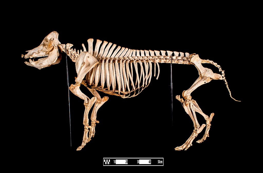
Afrikaans: 'n Varkskelet wat deur die proses van beenmaserasie voorberei is, en by die Museum vir Veeartseny-anatomie FMVZ USP uitgestal word. Varke is omnivore met 44 tande, insluitende hul geboë slagtande, wat by die beer voortdurend groei, maar nié by die sog nie. Hulle kort ledemate het vier tone elk en is met hoewe afgerond. Die kop het 'n driehoekige profiel wat eindig in die skyfagtige snoet, ondersteun deur die rostrale been wat met die neuskraakbeen verbind is. Hierdie anatomiese konfigurasie stel die vark in staat om sy neus as 'n skopgraaf aan te wend om wortels uit te grawe. Varkvleis is die mees verbruikte vleis ter wêreld, en verteenwoordig sowat 45% van die wêreldwye vleismark.
English: Swine. Sus
Skeleton specimen of a swine prepared by bone maceration technique in display at the Museum of Veterinary Anatomy FMVZ USP. Pigs are omnivores and have 44 teeth, including the curved canines or tusks, which are of continuous growth in the boar but not in the sow. They have short limbs with four fingers finishing in hoofs. The head has a triangular profile ending at the disclike snout, which is supported by the rostral bone attached to the nasal cartilages. This anatomical configuration of the muzzle allows the pig to use its nose as a shovel to dig roots. Pork is the most consumed meat in the world – corresponding to about 45 % of the global meat market. This file was published as the result of a partnership between the Museum of Veterinary Anatomy FMVZ USP, the RIDC NeuroMat and the Wikimedia Community User Group Brasil. This GLAM project is reported. Photography: Museum of Veterinary Anatomy FMVZ USP Author: Wagner Souza e SilvaEspañol: Esqueleto de cerdo después de la aplicación de una técnica de maceración ósea, en exhibición en el Museo de Anatomía Veterinaria de la Universidad de São Paulo, Brasil.
Polski: Szkielet świni spreparowany za pomocą techniki maceracji kości i wystawiony w Muzeum Anatomii Zwierząt Wydziału Weterynarii i Zootechniki Uniwersytetu w São Paulo (port. Museu de Anatomia Veterinária da Faculdade de Medicina Veterinária e Zootecnia da USP).
Português: Esqueleto de suíno, após técnica de maceração óssea, em exibição no Museu de Anatomia Veterinária da Universidade de São Paulo
Українська: Скелет свині, до якого було застосовано техніку мацерації кісток, на експозиції в Музеї ветеринарної анатомії Університету Сан-Паулу, Бразилія.
Čeština: Kostra prasete (svině) vystavená v Muzeu veterinární anatomie (FMVZ USP) Univerzity São Paulo, Brazílie.
Français : Squelette de porc, après une technique de macération osseuse; exposé au musée d'anatomie vétérinaire de l'université de São Paulo.
Magyar: Sertés csontváza a São Paulo Egyetem Állatorvosi Anatómiai Múzeumában
Italiano: Scheletro di suino, dopo la tecnica della macerazione ossea, in mostra al Museu de Anatomia Veterinária Prof. Dr. Plínio Pinto e Silva dell'Università di San Paolo.
한국어: 상파울루 대학교 수의해부학 박물관에 전시 중인 침연 과정을 거친 돼지의 뼈대.
Македонски: Скелет на свиња во Музејот на ветеринарна анатомија при Универзитетот во Сао Паоло, Бразил.
|
| 날짜 | |
| 출처 | Museum of Veterinary Anatomy FMVZ USP |
| 저자 | Museum of Veterinary Anatomy FMVZ USP / Wagner Souza e Silva |
| 다른 버전 |
.jpg)
수상
|

|
이 사진은 가치있는 사진 조건을 만족하며, 다음 분야에서 공용에서 가장 가치있는 사진으로 여겨집니다: Pig skeletons adult pig skeleton.. 이 사진에 관한 토론을 여기에서 보실 수 있습니다. |
이 이미지는 2017년 10월 1일의 오늘의 이미지로 선정되었습니다. 이미지 설명은 다음과 같습니다. 한국어: 상파울루 대학교 수의해부학 박물관에 전시 중인 침연 과정을 거친 돼지의 뼈대. 다른 언어들:
Čeština: Kostra prasete (svině) vystavená v Muzeu veterinární anatomie (FMVZ USP) Univerzity São Paulo, Brazílie. English: Swine skeleton, after technique of bone maceration, on display at the University of São Paulo Museum of Veterinary Anatomy. Español: Esqueleto de cerdo después de la aplicación de una técnica de maceración ósea, en exhibición en el Museo de Anatomía Veterinaria de la Universidad de São Paulo, Brasil. Français : Squelette de porc, après une technique de macération osseuse; exposé au musée d'anatomie vétérinaire de l'université de São Paulo. Italiano: Scheletro di suino, dopo la tecnica della macerazione ossea, in mostra al Museu de Anatomia Veterinária Prof. Dr. Plínio Pinto e Silva dell'Università di San Paolo. Magyar: Sertés csontváza a São Paulo Egyetem Állatorvosi Anatómiai Múzeumában Polski: Szkielet świni spreparowany za pomocą techniki maceracji kości i wystawiony w Muzeum Anatomii Zwierząt Wydziału Weterynarii i Zootechniki Uniwersytetu w São Paulo (port. Museu de Anatomia Veterinária da Faculdade de Medicina Veterinária e Zootecnia da USP). Português: Esqueleto de suíno, após técnica de maceração óssea, em exibição no Museu de Anatomia Veterinária da Universidade de São Paulo. Македонски: Скелет на свиња во Музејот на ветеринарна анатомија при Универзитетот во Саун Пауло, Бразил. Українська: Скелет свині, до якого було застосовано техніку мацерації кісток, на експозиції в Музеї ветеринарної анатомії Університету Сан-Паулу, Бразилія. 한국어: 상파울루 대학교 수의해부학 박물관에 전시 중인 침연 과정을 거친 돼지의 뼈대. |
라이선스
This media was produced by the Museum of Veterinary Anatomy (FMVZ USP) and was licensed as Creative Commons BY-SA 4.0. The MAV is an organ of integration of the School of Veterinary Medicine and Animal Science, University of São Paulo.
MAV-FMVZ USP asks to be cited as shown below. If the photographer name is mentioned, please, cite it after the museum's name. If not, just provide the reference to the museum. Attribution in English: Museum of Veterinary Anatomy FMVZ USP / name of the photographer when stated Attribution in Portuguese: Museu de Anatomia Veterinária da FMVZ USP / nome do fotógrafo quando atribuído |
- 이용자는 다음의 권리를 갖습니다:
- 공유 및 이용 – 저작물의 복제, 배포, 전시, 공연 및 공중송신
- 재창작 – 저작물의 개작, 수정, 2차적저작물 창작
- 다음과 같은 조건을 따라야 합니다:
- 저작자표시 – 적절한 저작자 표시를 제공하고, 라이센스에 대한 링크를 제공하고, 변경사항이 있는지를 표시해야 합니다. 당신은 합리적인 방식으로 표시할 수 있지만, 어떤 방식으로든 사용권 허가자가 당신 또는 당신의 사용을 지지하는 방식으로 표시할 수 없습니다.
- 동일조건변경허락 – 만약 당신이 이 저작물을 리믹스 또는 변형하거나 이 저작물을 기반으로 제작하는 경우, 당신은 당신의 기여물을 원저작물과 동일하거나 호환 가능한 라이선스에 따라 배포하여야 합니다.
Serkan yetişkin PTT25100 ustayetiskin@gmail.com
[[File:Brasq8ri6t|baş parmak|alt=Windows XP All DC LG |www.Google.com
파일 역사
날짜/시간 링크를 클릭하면 해당 시간의 파일을 볼 수 있습니다.
| 날짜/시간 | 섬네일 | 크기 | 사용자 | 설명 | |
|---|---|---|---|---|---|
| 현재 | 2016년 8월 11일 (목) 17:03 | | 3,923 × 2,592 (2.2 MB) | Rodrigo.Argenton | cleaning the background, increase light, sharpness, metadata... |
| 2016년 7월 27일 (수) 02:21 |  | 3,872 × 2,592 (1.81 MB) | Sturm | User created page with UploadWizard |
이 파일을 사용하는 문서
다음 문서 1개가 이 파일을 사용하고 있습니다:
이 파일을 사용하고 있는 모든 위키의 문서 목록
다음 위키에서 이 파일을 사용하고 있습니다:
- be-tarask.wikipedia.org에서 이 파일을 사용하고 있는 문서 목록
- crh.wikipedia.org에서 이 파일을 사용하고 있는 문서 목록
- cv.wikipedia.org에서 이 파일을 사용하고 있는 문서 목록
- cy.wikipedia.org에서 이 파일을 사용하고 있는 문서 목록
- de.wikipedia.org에서 이 파일을 사용하고 있는 문서 목록
- en.wikipedia.org에서 이 파일을 사용하고 있는 문서 목록
- fa.wikipedia.org에서 이 파일을 사용하고 있는 문서 목록
- fr.wikipedia.org에서 이 파일을 사용하고 있는 문서 목록
- hu.wikipedia.org에서 이 파일을 사용하고 있는 문서 목록
- hy.wikipedia.org에서 이 파일을 사용하고 있는 문서 목록
- incubator.wikimedia.org에서 이 파일을 사용하고 있는 문서 목록
- it.wiktionary.org에서 이 파일을 사용하고 있는 문서 목록
- ka.wikipedia.org에서 이 파일을 사용하고 있는 문서 목록
- krc.wikipedia.org에서 이 파일을 사용하고 있는 문서 목록
- lbe.wikipedia.org에서 이 파일을 사용하고 있는 문서 목록
- lez.wikipedia.org에서 이 파일을 사용하고 있는 문서 목록
- mdf.wikipedia.org에서 이 파일을 사용하고 있는 문서 목록
- mg.wikipedia.org에서 이 파일을 사용하고 있는 문서 목록
- mk.wikipedia.org에서 이 파일을 사용하고 있는 문서 목록
- os.wikipedia.org에서 이 파일을 사용하고 있는 문서 목록
- outreach.wikimedia.org에서 이 파일을 사용하고 있는 문서 목록
- pt.wikipedia.org에서 이 파일을 사용하고 있는 문서 목록
- ru.wikipedia.org에서 이 파일을 사용하고 있는 문서 목록
- ru.wikinews.org에서 이 파일을 사용하고 있는 문서 목록
- sah.wikipedia.org에서 이 파일을 사용하고 있는 문서 목록
- sv.wikipedia.org에서 이 파일을 사용하고 있는 문서 목록
- tt.wikipedia.org에서 이 파일을 사용하고 있는 문서 목록
- tyv.wikipedia.org에서 이 파일을 사용하고 있는 문서 목록
- udm.wikipedia.org에서 이 파일을 사용하고 있는 문서 목록
- uk.wikipedia.org에서 이 파일을 사용하고 있는 문서 목록
- vep.wikipedia.org에서 이 파일을 사용하고 있는 문서 목록
- xal.wikipedia.org에서 이 파일을 사용하고 있는 문서 목록
- zh.wikipedia.org에서 이 파일을 사용하고 있는 문서 목록